Schaffer-Nishimura lab talk for Cornell Engineering

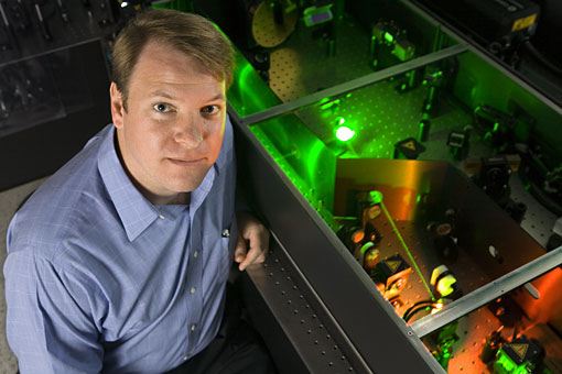
Femtosecond laser pulses have the unique capability to deposit energy into a microscopic volume in the bulk of a transparent material without affecting the surface of the material. Here we propose to develop and use this capability to disrupt specifically targeted structures in live cells and animals with the goal of elucidating function and modeling disease states. As work to optimize laser parameters, beam delivery methods, and pulse manipulation schemes to achieve the best possible precision and penetration. In parallel with this technology development, our studies focus on the use of femtosecond laser disruption to answer pressing scientific questions in several areas, including development of methods for targeted gene transfection through laser poration of cell membranes, determination of the neural wiring that encodes behavior in small animals, and the production of animal models of cerebrovascular diseases. Critically, studies in this collaboration will span biological scales, from disruption of sub-cellular structures in cell culture, to in vivo ablation of cellular and sub-cellular features in small organisms and mammals, to pre-clinical evaluation of laser-based therapeutic strategies of relevance for human disease. This work is carried out by a team that includes experts on femtosecond laser physics and laser/material interactions as well as experts on cell biology, neurobiology, and pathology. This broad approach to the development of femtosecond laser disruption will allow us to fully explore the capabilities and limitations of this technology as a tool to fill the scientific need for precise, targeted physical disruption of biological structures.

My interest is in studying the contribution of multiple physiological systems to disease initiation and progression, with applications in neurodegenerative disease, cardiovascular disease, and cancer. We would like to understand how the vascular, immune, inflammatory systems and cells native to a tissue interact in these diseases. A major challenge in such work is that model systems such as cell culture or even organotypic tissue culture cannot fully recapitulate all the different cell types involved in disease, so in vivo studies are required. However, it is experimentally difficult to study and manipulate cell-level dynamics in live animals. Recently, we have worked to develop technologies such as improved imaging using multiphoton microscopy that work in whole animals and have sufficient spatial and temporal resolution to quantify cellular dynamics. We also now have tools, femtosecond laser ablation, to produce targeted disruption with cellular-scale precision. We used these tools to piece together a picture of how occlusion or hemorrhage of small blood vessels in the brain affects the health and function of nearby neurons, and thus contributes to cognitive decline. The goal of our work is to continue to use such capabilities to unravel the interaction of various physiological systems in diseases, with special interests in Alzheimer’s disease, cancer metastasis, and cardiac blood flow.
We are grateful to the following private and public agencies for the support of our research.